
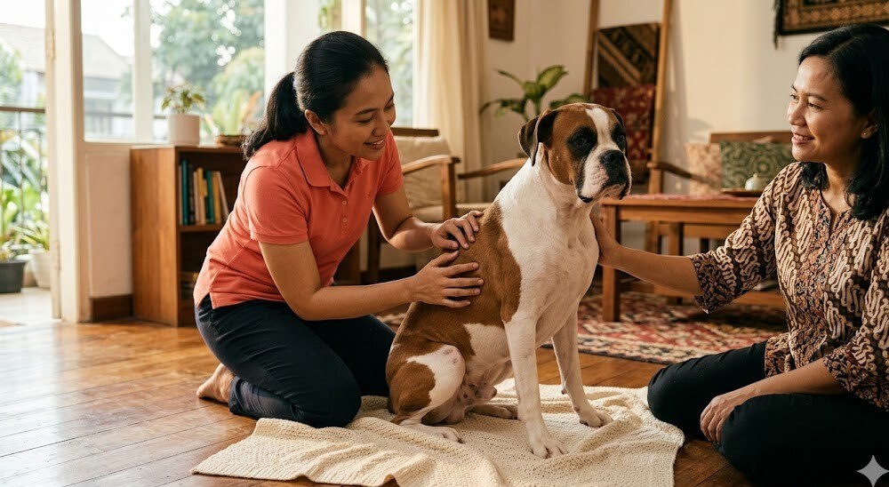
"Doc, my 8-year-old Boxer has a new lump on the side of his body. It was small at first but now it's red and sometimes itchy. When I felt it, it actually got redder and more swollen — is this a malignant tumor?" This question comes into our WhatsApp often, and for Boxers in particular our answer is always: don't wait any longer — every new skin lump in a dog, especially one whose behavior changes when palpated, must have an FNA before anything else. Mast cell tumor is the most common skin tumor in dogs and Boxers have a very high predisposition.
This article is a complete guide for owners who have just found a skin lump, owners of predisposed breeds (Boxer, Pug, Bulldog, Boston Terrier, Labrador, Golden Retriever), or owners who already have an MCT diagnosis and are weighing treatment options and prognosis.
What a mast cell tumor is and why dogs get it most often
A mast cell tumor (MCT, mastocytoma) is a tumor of the mast cell — an immune cell that normally plays a role in allergic and inflammatory reactions. Mast cells normally reside in connective tissue in the skin, respiratory mucosa, and gastrointestinal tract. In MCT, these cells become neoplastic and form a tumor — most often in the skin (cutaneous MCT), sometimes in internal organs (visceral MCT, more rare).
An important point to underline: MCT is the most common skin tumor in dogs — the figure often cited in the international oncology literature is roughly 16-21% of all canine skin tumors (London & Thamm, Withrow & MacEwen Small Animal Clinical Oncology 6e). In daily practice, every new skin lump in a dog must be suspected as MCT until proven otherwise.
Predisposed breeds
Several breeds have a significantly higher MCT risk than the general dog population. Per the ACVIM Oncology Consensus Statement on MCT and Withrow & MacEwen 6e, predisposed breeds include:
- Boxer — highest risk, roughly 3-8× that of the general population. Almost every Boxer over 6 years old needs routine screening for skin lumps
- Pug — often multiple MCTs, many of them low grade
- Bulldog (English Bulldog, French Bulldog)
- Boston Terrier
- Labrador Retriever
- Golden Retriever
- Beagle
- Shar-Pei — often appears younger and higher grade (a clinically aggressive form)
- Weimaraner, Rhodesian Ridgeback, Cocker Spaniel also appear on the list
Age distribution — most MCTs appear in dogs 8-10 years old, but they can also appear young (4-6 years), especially in breeds like the Shar-Pei.
Pathogenesis — why a mast cell tumor is "different" from an ordinary skin tumor
This is what makes MCT unique and requires careful handling: mast cells contain granules full of vasoactive substances:
- Histamine — causes itching, redness, vasodilation, and if released massively can cause anaphylactic shock
- Heparin — an anticoagulant, can cause a local bleeding tendency if released massively
- Proteolytic enzymes (tryptase, chymase) — tissue destruction, edema
- Cytokines and chemokines — inflammatory mediators
Important clinical implications:
- "Darier sign" — when an MCT is palpated or manipulated, granules can be released locally, producing a red flare, swelling, and itching around the tumor. This is a characteristic sign owners often notice ("Every time I touch it, it turns red and swollen"). This is also why an MCT must not be manipulated carelessly
- Risk of systemic degranulation during handling/surgery — a massive release during surgery can cause intraoperative hypotension, GI ulceration (histamine-mediated), and a bleeding tendency. Because of this, pre-medication with antihistamines (H1 + H2 blockers) before FNA and especially before surgery is the standard of care
- Systemic GI ulceration — in large or metastatic MCTs, systemic histamine can cause gastric ulceration (vomiting with blood, melena). For this reason omeprazole or famotidine is often added to the regimen
- Coagulation issues — local heparin can cause more bleeding than expected during surgery
Key message for owners: do not keep squeezing, touching, or "exploring" a lump suspected to be MCT — repeated palpation can trigger degranulation. See the vet, and let the vet assess it carefully.
Clinical signs — what owners notice
MCT has a "great masquerader" reputation — its appearance is highly variable, able to mimic benign tumors (lipoma, histiocytoma, papilloma) or a skin infection. For this reason it cannot be judged visually alone — an FNA is mandatory for every new skin lump in a dog.
The spectrum of MCT appearance often encountered:
- Raised cutaneous nodule — can be small (a few mm) up to large (several cm), single or multiple
- Variable consistency — can be firm, soft, or elastic
- Variable color — can be pink, reddish, purple, or normal skin color
- Variable surface — can be smooth (like a lipoma), rough/papillomatous, or with hair loss over it
- Ulceration — in more advanced or aggressive MCTs, there is often an open ulcer on the surface
- Positive Darier sign — when palpated, it becomes acutely red-swollen-itchy. This is a highly suggestive sign of MCT
- Variable growth — can be slow and stable, can grow quickly, or can fluctuate in size (enlarging during degranulation, shrinking when quiet)
- Itching and irritation — sometimes the dog licks or scratches the tumor area because of histamine release
Most common locations: the trunk and the extremities (legs) are the most common sites. Inguinal, perineal, and mucosal locations (oral, prepuce, vulva) were historically considered "high-risk sites" with worse prognosis, although modern data show grade is more important than location.
Because of this variability of appearance → PRACTICAL RULE: every new skin lump in a dog ≥1cm MUST have an FNA first before anything else (before being observed at home, before being scheduled for routine surgery, before a steroid injection). "Let's wait, check again in 2 weeks" for a skin lump in a dog is now an outdated practice.
Diagnosis — FNA first, then biopsy
Step 1: Thorough physical examination
- Inspect all skin and mucosa — MCT can be multiple, check the whole body
- Palpate carefully (not too aggressively) — note size, consistency, mobility, Darier sign
- Palpate all regional lymph nodes (nearest to the tumor and along the drainage path)
- Body condition score, measure the tumor, document with photos and calipers
Step 2: Pre-medication before FNA
Giving an antihistamine before FNA in a dog with suspected MCT is standard practice per Plumb's 7e + ACVIM Oncology Consensus on MCT:
- Diphenhydramine (H1 antihistamine) — given IM/SC before FNA or tumor manipulation
- Famotidine or ranitidine (H2 antihistamine) — for protection against gastric ulceration
- Dosing per Plumb's 7e — must be administered by a veterinarian, not by the owner
Step 3: FNA cytology — the first-tier diagnosis
- FNA from the primary mass and from the regional lymph nodes
- MCT cytology is highly distinctive — a monomorphic mast cell population with characteristic cytoplasmic granules (a round cell tumor with metachromatic granules on Diff-Quick or Wright-Giemsa staining)
- FNA is often enough to diagnose "this is MCT" — but not enough for grading
- FNA of regional lymph nodes — assesses spread to the nodes
Step 4: Staging work-up
After the MCT diagnosis is confirmed via FNA, staging is done to determine the extent of disease and plan treatment:
- FNA of all regional lymph nodes — even if they do not feel enlarged (normal size ≠ no metastasis). The primary drainage node must be FNA'd
- Abdominal ultrasound — check the spleen and liver (the most common visceral metastasis targets). The spleen/liver can also be FNA'd during the ultrasound if there is a suspicious lesion
- CBC with buffy coat smear — check for peripheral mastocytemia (mast cells circulating in the blood, suggestive of advanced disease)
- Biochemistry + urinalysis — baseline organ function pre-treatment
- Thoracic radiographs — less common as MCT metastasis, but useful for pre-anesthetic screening plus a baseline if there is concern
- Bone marrow aspirate — usually for advanced cases or peripheral mastocytemia
Step 5: Histopathology biopsy — the gold standard for grading
After surgery (or a pre-surgery incisional biopsy for planning), the specimen must be sent to a pathologist for grading. Grading is the MOST IMPORTANT prognostic factor and determines the subsequent treatment plan.
MCT grading — Patnaik + Kiupel
There are two grading systems used in parallel in modern practice:
Patnaik (1984) — 3-tier system
- Grade I (well-differentiated) — mature mast cells in the superficial dermis, mitotic figures rare, very good prognosis. ~25% of cases
- Grade II (intermediately-differentiated) — the largest category (~50% of MCTs) but a highly heterogeneous prognosis. This is the "grey zone" with limited predictivity — some behave indolently, some metastasize quickly. Because of this, the Kiupel 2-tier system was developed
- Grade III (poorly-differentiated) — anaplastic cells, high mitotic figures, frequent ulceration, often invasive into the subcutis and muscle, poor prognosis with high metastasis. ~25% of cases
Kiupel (2011) — 2-tier system
- Low-grade — low mitotic count, no karyomegaly, no multinucleated cells, no atypical nuclei. Good prognosis, many curative with surgery alone
- High-grade — mitotic count ≥7 per 10 high-power fields, karyomegaly, multinucleated cells, atypical nuclei. Poor prognosis with high metastasis
Most modern pathologists report both systems in parallel (Patnaik + Kiupel) for maximum prognostic information. The combination of grading + location + staging + c-kit mutation status determines the treatment plan.
c-kit mutation status (KIT proto-oncogene)
Some MCTs have a mutation in the KIT gene (c-kit) — a proto-oncogene important in mast cells. MCTs with a KIT mutation (about 15-30% of all MCTs, higher in high-grade) tend to be more aggressive but are also responsive to targeted therapy (toceranib/Palladia). Testing for c-kit mutation at a specialist lab can guide treatment selection.
Treatment — surgery first, then evaluate
Pillar 1: Wide surgical excision
Surgery is the primary and most important treatment for MCT, especially grade I-II low-grade. The principles:
- Wide margin of 2-3cm laterally and 1 fascial plane deep — the historical standard. Modern studies show a 2cm lateral margin + 1 fascial plane deep is often adequate for grade I-II, but 3cm for grade III or high-grade Kiupel
- Histopathologic margin assessment — the pathologist evaluates whether the margins are "clean" (no tumor cells at the edge). Non-clean margins = risk of local recurrence
- Re-excision or radiation if the margins are not clean — discuss with an oncologist
- Sentinel lymph node biopsy — increasingly recommended for accurate staging, especially for grade II-III
Pillar 2: Adjuvant chemotherapy (for grade III high-grade or metastatic)
For grade III MCT, high-grade Kiupel, with lymph node metastasis, or non-clean margins where radiation is not available, adjuvant systemic chemotherapy is recommended. Per Plumb's 7e + ACVIM Oncology Consensus on MCT, the drugs often used:
- Vinblastine + prednisone — a combination often used as first-line adjuvant
- Lomustine (CCNU) + prednisone — an alternative or rescue protocol
- Cyclophosphamide — sometimes added to the protocol
- Commitment: a series of visits for drug administration plus CBC monitoring
Pillar 3: Targeted therapy — toceranib (Palladia)
Toceranib phosphate (Palladia) is a tyrosine kinase inhibitor that targets the KIT receptor. Approved for MCT in dogs (FDA-approved 2009, available in many countries including Indonesia via specialist clinics).
- Particularly effective for MCT with a c-kit mutation
- Given orally at home (tablet)
- Side effects can be significant: GI upset, neutropenia, proteinuria, hypertension — routine monitoring is needed
- Discuss with an oncologist to evaluate candidacy plus a monitoring plan
- Per Plumb's 7e for dosing and the monitoring protocol
Pillar 4: Supportive — H1 + H2 blockers
- Diphenhydramine or cetirizine — H1 blocker
- Famotidine or omeprazole — H2 blocker for gastric ulceration protection
- Given routinely especially during the treatment phase and if there is a significant tumor burden
Pillar 5: Radiation therapy (rare in Indonesia)
Adjuvant radiation is an option for non-clean margins plus a location that cannot be re-excised widely. But veterinary radiation oncology infrastructure is still limited in Indonesia — this option is rarely available. Discuss with an oncologist.
Prognosis — honest about expectations per grade
| Category | Outcome with Adequate Treatment |
|---|---|
| Patnaik Grade I / Kiupel Low-grade, clean margins | Mostly curative with surgery alone. Long-term survival is very good (>2 years, many live normally to the end of their lifespan) |
| Patnaik Grade II / Kiupel Low-grade, clean margins | Mostly favorable — median survival >2 years for many cases, but heterogeneous |
| Patnaik Grade II / Kiupel High-grade | Variable — needs close monitoring plus consideration of adjuvant therapy. Median survival 1-2 years |
| Patnaik Grade III / Kiupel High-grade | Poor — high metastatic risk, median survival often <1 year even with multimodal treatment. Some cases respond to toceranib (especially c-kit mutated) |
| Metastasis to lymph node | Prognosis declines significantly; adjuvant systemic therapy is needed |
| Metastasis to viscera (spleen, liver, bone marrow) | Poor — survival mostly in months |
Favorable prognostic factors: low-grade (Kiupel), low Patnaik grade, low mitotic index, clean surgical margins, no nodal involvement, no c-kit mutation, non-high-risk location, single tumor.
Unfavorable prognostic factors: high-grade Kiupel, Patnaik Grade III, high mitotic index, dirty margins, nodal/visceral metastasis, c-kit mutation (without targeted therapy), peripheral mastocytemia, ulcerated/recurrent tumor, inguinal/perineal/mucosal location (historically), multiple tumors.
Key message for owners — when to see the vet
Practical rules that owners of predisposed breeds (Boxer, Pug, Bulldog, Boston, Labrador, Golden, Beagle, Shar-Pei) should hold on to:
- Every new skin lump in a dog ≥1cm MUST have an FNA — it must not be observed at home, must not be "wait 2 weeks to see if it changes." FNA is simple, fast, with results in 1-3 days
- A lump with a positive Darier sign (red-swollen-itchy when palpated) → urgent FNA
- A lump that grows quickly (clearly changing size within weeks) → urgent FNA
- An ulcerated lump (an open wound on the surface) → urgent FNA + staging
- Multiple skin nodules in a predisposed breed → FNA all of them, MCT can be multiple
- A dog with a history of MCT → routine screening at every visit; the owner should palpate routinely at home (gently, not constantly)
- Do not keep squeezing/touching a lump suspected to be MCT — repeated palpation can trigger degranulation with local/systemic side effects
- Do not inject steroid directly into a dog's skin lump without an FNA first — steroids can mask the symptoms of MCT, delay diagnosis, and compromise treatment
Canine Mast Cell Tumor FAQ
My dog is a Boxer with a small skin lump — is it definitely MCT?
Not definitely, but Boxers have a significantly higher MCT risk than other breeds. It cannot be determined visually alone — it could be MCT, a lipoma, a histiocytoma (a benign tumor that often appears in young dogs and resolves on its own), a cyst, or another tumor. FNA is mandatory to confirm. FNA is simple, results come in 1-3 days, and it immediately guides the treatment plan. Do not delay.
Is a mast cell tumor contagious to other dogs or to humans?
No. MCT is the dog's own cancer cells — it is not contagious to other dogs, cats, or humans. No quarantine is required.
My dog has had MCT surgery, when should the follow-up be?
It depends on grade + margins + staging. A common rule of thumb: a clinical check + palpation (especially of the surgical scar and regional lymph nodes) every 1-3 months in the first year, then every 6 months. For grade III or high-grade — re-staging with abdominal ultrasound + FNA of regional nodes every 3 months. Discuss the specific schedule with the veterinarian based on your dog's risk profile.
How much does MCT treatment cost in Indonesia?
It varies greatly depending on the type of treatment and clinic. Wide excision surgery at a regular surgical clinic is relatively affordable. Adjuvant chemotherapy (vinblastine/lomustine) or toceranib (Palladia) is significantly more expensive — given at a specialist clinic. Discuss specific estimates with your veterinarian or a referral oncology clinic. Ask Prabasavet on WhatsApp if you need a referral to the nearest oncology service in your area.
Can my dog have anaphylaxis during or after MCT surgery?
There is a risk (systemic degranulation during manipulation of a large tumor), but with proper pre-medication (H1 + H2 antihistamines before and during anesthesia) + intraoperative monitoring + careful tumor handling, the risk of anaphylaxis can be significantly mitigated. It is not a reason to delay surgery. Make sure your surgeon is aware that the tumor is an MCT and applies the standard pre-medication protocol.
My dog has grade III high-grade MCT — is toceranib (Palladia) worth it?
Discuss with an oncologist. Toceranib is particularly effective for MCT with a c-kit mutation (testing is available at a specialist lab). For high-grade cases with a c-kit mutation, toceranib shows a significant response rate plus an extension of quality of life. Side effects can be significant (GI upset, neutropenia, proteinuria) so routine monitoring is needed. Cost and the monitoring commitment also need to be considered. Not all high-grade MCTs respond — the decision is based on your dog's specific profile.
Can I buy OTC antihistamines for my dog with MCT on my own?
Not without consulting a veterinarian first. Diphenhydramine, cetirizine, and famotidine are indeed commonly prescribed, but the dosing must be adjusted for body weight + interval + duration by the veterinarian. Self-medication without proper diagnosis + staging can mask symptoms and delay diagnosis. See the veterinarian for a complete plan, then prescribed medication can be continued at home under supervision.
My dog is a senior >10 years old with a new MCT — can it still have surgery?
Age is not the only factor. What matters: the dog's general condition, comorbidities (heart, kidney, liver), the tumor profile (grade, location, staging), and owner preference. Many senior dogs tolerate surgery well with an adequate pre-op work-up (blood work + ECG + BP) + an anesthesia protocol appropriate for geriatrics. Discuss with the veterinarian based on your dog's specific condition, not age alone.
After MCT surgery, can my dog return to normal activity?
Most cases, yes — especially for grade I-II low-grade with clean margins. Recovery from surgery is generally 2-3 weeks (wound healing), after which normal activity can resume. For cases with adjuvant chemotherapy or toceranib, there are lifestyle adjustments during the treatment phase (avoid excess stress, monitor side effects, etc.). Discuss the rehabilitation and lifestyle plan with the veterinarian.
Summary
Mast cell tumor is the most common skin tumor in dogs (about 16-21% of all canine skin tumors). Significant predisposed breeds: Boxer, Pug, Bulldog, Boston Terrier, Labrador, Golden Retriever, Beagle, Shar-Pei. What makes MCT unique and requires careful handling: mast cells contain granules of histamine + heparin + enzymes, so manipulating the tumor can trigger degranulation with local (Darier sign) or systemic (anaphylaxis, GI ulceration, bleeding) side effects.
MCT appearance is highly variable — so the practical rule: every new skin lump in a dog ≥1cm MUST have an FNA first, it must not be observed at home or "wait 2 weeks." Diagnosis via FNA cytology + staging (FNA of regional lymph nodes + abdominal ultrasound + CBC buffy coat) + histopathology biopsy for grading.
Grading is the most important prognostic factor: Patnaik I-II-III + Kiupel low/high-grade. Wide excision surgery (margin 2-3cm + 1 fascial plane deep) is the primary treatment — most grade I-II low-grade are curative with surgery alone. For grade III or high-grade Kiupel or metastatic: adjuvant chemotherapy (vinblastine, lomustine) or targeted therapy toceranib (Palladia) for c-kit mutated. Pre-medication with H1 + H2 antihistamines before FNA and surgery is standard.
Prognosis: low-grade with clean margins is mostly curative. High-grade with metastasis has a poor prognosis — a realistic discussion with an oncologist + a multimodal plan is very important. Do not keep squeezing/touching a lump suspected to be MCT, do not inject steroid directly without an FNA first.
Does your predisposed-breed dog have a new skin lump and need an FNA without the stress of a trip to a crowded clinic? See Prabasavet's pet care guide, the pet emergency guide, or contact us on WhatsApp for a free consultation and a referral to the nearest oncology service in your area.
Read also: Dogs Afraid of the Clinic: The House Call Solution, Dog Vomiting Blood: Causes and Emergency Management.
Medical references used in this article
This article was prepared with reference to the following sources, verified per clinical sentence:
- Vail DM, Thamm DH, Liptak JM (editors). Withrow and MacEwen's Small Animal Clinical Oncology, 6th edition — canine mast cell tumor chapter (epidemiology, breed predisposition, pathogenesis of histamine/heparin granules, clinical signs, staging, Patnaik + Kiupel grading, surgical margin recommendations, adjuvant chemotherapy, prognosis per grade)
- ACVIM (American College of Veterinary Internal Medicine) Consensus Statement on Mast Cell Tumours in Dogs — standard diagnostic protocol, pre-medication recommendations, staging work-up, surgical principles, adjuvant therapy decision algorithm
- Patnaik AK, Ehler WJ, MacEwen EG. Canine cutaneous mast cell tumor: morphologic grading and survival time in 83 dogs. Veterinary Pathology 1984 — the original 3-tier Patnaik grading system, correlation of grade with survival
- Kiupel M, Webster JD, Bailey KL, et al. Proposal of a 2-tier histologic grading system for canine cutaneous mast cell tumors to more accurately predict biological behavior. Veterinary Pathology 2011 — the modern 2-tier Kiupel grading system with better predictivity for the Patnaik II "grey zone"
- London CA, Thamm DH. Mast Cell Tumors chapter in Withrow & MacEwen — a comprehensive overview of diagnosis-staging-treatment-prognosis
- Plumb's Veterinary Drug Handbook, 7th edition — monographs for: diphenhydramine (H1 antihistamine pre-FNA + surgery), famotidine + omeprazole (H2 + PPI GI protection), vinblastine + lomustine (CCNU) + cyclophosphamide (adjuvant chemotherapy for MCT), prednisone (palliative + adjuvant), toceranib phosphate Palladia (tyrosine kinase inhibitor for c-kit mutated MCT)
- London CA, Malpas PB, Wood-Follis SL, et al. Multi-center, placebo-controlled, double-blind, randomized study of oral toceranib phosphate (SU11654), a receptor tyrosine kinase inhibitor, for the treatment of dogs with recurrent (either local or distant) mast cell tumor following surgical excision. Clinical Cancer Research — landmark trial on toceranib efficacy in recurrent/metastatic MCT
- Webster JD, Yuzbasiyan-Gurkan V, Kaneene JB, et al. The role of c-KIT in tumorigenesis: evaluation in canine cutaneous mast cell tumors. Neoplasia 2006 — c-kit mutation prevalence + correlation with prognosis + response to targeted therapy
This article is general guidance based on international guidelines (ACVIM) and standard oncology literature. For your dog's specific condition — including tumor grade, staging, c-kit mutation status, comorbidities, and treatment preferences — consulting a veterinarian and/or referral to a specialist oncologist is the right step. Antihistamine pre-medication before FNA or MCT surgery, and chemotherapy/targeted therapy protocols, must be administered and monitored by a veterinarian competent in veterinary oncology; self-administration is not recommended.


