"My German Shepherd is 7 months old, and when he runs in the park his movement looks odd — his hind legs often jump together like a rabbit, not a normal run. When he gets up from sleeping he looks stiff, and he often stops midway when taken on a long walk. Is this normal for a GSD puppy, or a sign of hip dysplasia?" This question comes up often to the Prabasavet team from large breed puppy owners. The answer in most cases: an orthopedic evaluation + radiograph is needed, because a "bunny-hopping" gait in a large breed puppy is one of the classic signs of hip dysplasia.
Hip dysplasia is the most common hereditary orthopedic disorder in large and giant breed dogs. Its presentation varies — from a 4-12 month-old puppy starting to limp, to an adult over 2 years old who only shows signs because of the secondary osteoarthritis that gradually develops over time.
This article helps you recognize the early signs of hip dysplasia, understand how a vet diagnoses it via OFA radiograph and PennHIP, and learn the non-surgical and surgical management options relevant to your dog's condition.
What is hip dysplasia in dogs?
Hip dysplasia (coxofemoral dysplasia) is a developmental malformation of the hip joint in which joint laxity (looseness of the joint) causes the femoral head (the head of the thigh bone) to not sit stably in the acetabulum (the hip socket). As a result of this chronic instability, the joint undergoes progressive damage: repeated subluxation, cartilage micro-trauma, synovial inflammation, bone remodeling, and ultimately secondary osteoarthritis.
This disease has two main pathophysiology pathways:
- Genetic component — hip dysplasia is a polygenic disease with moderate-to-high heritability. Dogs from a lineage with hip dysplasia have a much higher risk, which is why OFA (Orthopedic Foundation for Animals) recommends that breeders screen the parents' hips before breeding.
- Environmental factors that aggravate it — overly rapid growth in a large breed puppy (over-feeding or a high-calorie diet), being overweight, high-impact exercise while the bones are still growing, and slippery surfaces that cause repeated slips can accelerate the expression of hip dysplasia even in a dog with moderate genetic risk.
The end result is almost always the same: progressive osteoarthritis (OA) in the hip joint. That is why hip dysplasia is often discussed together with osteoarthritis in senior dogs — hip dysplasia is one of the main causes of hip OA in adult dogs, and managing secondary OA becomes the pillar of long-term care after a hip dysplasia diagnosis.
Predisposed breeds — why large and giant breeds are most prone
Hip dysplasia can occur in any breed, including small breeds and mixed breeds, but the prevalence is highest in large and giant breeds. The breeds most often identified as predisposed based on OFA population data:
- German Shepherd — the breed most often associated with hip dysplasia in the public's mind, with a high radiographic prevalence in non-screened populations
- Labrador Retriever — popular and widely kept; the combination of popularity + predisposition means many clinical cases come from this breed
- Golden Retriever — often comorbid with elbow dysplasia too, needing screening of both joints
- Rottweiler — predisposition to hip + elbow dysplasia + cardiac issues, a breed that needs early orthopedic screening
- Saint Bernard and Newfoundland — giant breeds with very high mechanical load on the joints, often showing signs earlier
- Great Dane, Bernese Mountain Dog, Mastiff — other giant breeds that consistently make the predisposition list
- Bulldog — although not a large breed, its hip anatomy is highly predisposing
Mixed-breed dogs with a large breed component can also be affected. The important thing to emphasize: having a predisposed breed does not mean hip dysplasia is certain — most dogs of the breeds above that come from OFA-cleared parents and are raised with good weight + exercise management do not develop symptomatic hip dysplasia. But high awareness is important for early screening.
Clinical signs of hip dysplasia — distinguishing juvenile vs adult
Hip dysplasia has two different clinical presentations based on the age of onset, and the signs that appear also differ slightly between the two.
Juvenile form (4-12 months) — joint laxity dominant
In a growing large breed puppy, signs appear because of joint laxity and repeated subluxation. Characteristics:
- Bunny-hopping gait — when running, both hind legs jump together like a rabbit, rather than moving in alternation. This is a very characteristic sign of juvenile hip dysplasia and is often the first signal owners notice
- Intermittent lameness in the hind legs — sometimes appearing after exercise, disappearing after rest, then reappearing
- Reluctance to jump — a dog that should be energetic suddenly hesitates to climb stairs, get into the car, or jump onto the sofa
- Pelvic limb weakness — the hind legs appear weak, the dog often sits in a "frog" position (hind legs splayed out to the side)
- Stopping repeatedly while walking — energy seems less consistent than same-breed peers
- Reluctance to play rough or chase a ball as far as before
Adult form (>2 years) — secondary osteoarthritis dominant
In an adult dog, the signs relate to degenerative OA that has developed as a consequence of chronic joint laxity since youth. Many dogs with hip dysplasia show no clear signs until adulthood because the body compensates over the years. Characteristics:
- More consistent or permanent lameness in one or both hind legs
- Stiffness after a long rest — stiff for the first 5-10 minutes after getting up, then "warming up" and moving more smoothly
- Gluteal and thigh muscle atrophy — the muscles of the hip and rear thigh shrink due to chronic underuse, and the hind legs look thinner than the front legs
- Reluctance to climb stairs or refusal to go down
- A different way of getting up — the dog uses the front legs first then "drags" the rear body
- Decreased overall activity, a calmer mood, sometimes irritable when the hip area is touched
- In severe cases: the dog has difficulty standing without help, frequently falls, or refuses to walk more than a few meters
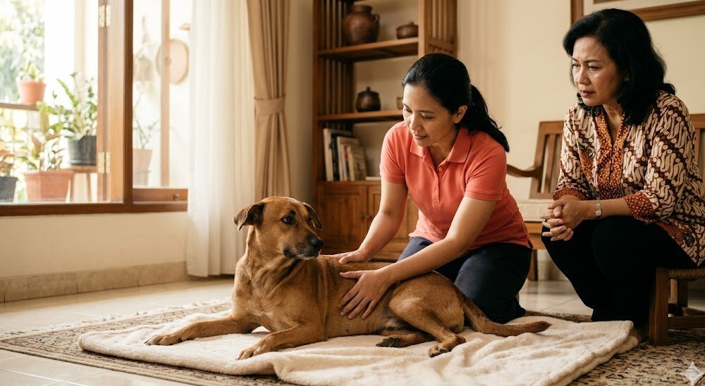
If you notice a cluster of the signs above in a large breed puppy or an adult dog, do not wait until it is severe. An orthopedic evaluation + radiograph can confirm the diagnosis and the grade. For owners in Jakarta and Greater Jakarta who find it difficult to bring a large, already-painful dog to a clinic (dogs with severe hip dysplasia often experience added stress and pain when getting into a car), WhatsApp Prabasavet for an initial discussion.
How a vet diagnoses hip dysplasia
History and orthopedic examination
The vet starts with a detailed history: breed, age of onset, symptom pattern (worse during exercise? worse after rest?), daily activity, what the dog ate during the growing phase, and the parents' hip history if from a breeder. Then a physical examination with an orthopedic focus:
- Observe the gait on a flat floor and while climbing/descending stairs or a curb — note bunny-hopping, asymmetry, or short stride
- Palpate the hip — check for swelling, local warmth, pain on manipulation, range of motion
- Check gluteal and thigh muscle symmetry — unilateral or bilateral atrophy
- Ortolani sign — a test characteristic of hip dysplasia. The dog is positioned in lateral or dorsal recumbency, and the vet manipulates the hip to detect a subluxation-reduction palpable as a "click" or "snap". A positive Ortolani is highly suggestive of hip dysplasia, especially in puppies
- Other orthopedic tests (drawer test to eliminate CCL rupture, manipulation of surrounding joints for differential diagnosis)
Standard radiograph (OFA hip evaluation)
The baseline imaging for evaluating hip dysplasia. What is seen on the radiograph: the shape of the acetabulum, the depth of femoral head coverage, joint space symmetry, and signs of secondary OA (osteophytes, subchondral sclerosis, joint capsule calcification).
OFA (Orthopedic Foundation for Animals) hip evaluation is the radiographic screening standard developed for breeding stock dogs in America. The standard OFA position is the ventrodorsal hip-extended view, which usually requires sedation or brief anesthesia so the dog relaxes and the position is accurate. OFA evaluation is performed at 24 months of age and above, because before that the bone and joint maturity is not final and the result can be over-optimistic.
The OFA grading scheme has 7 categories from "Excellent → Good → Fair" (normal range) to "Borderline → Mild → Moderate → Severe" (dysplastic range). OFA results serve as a global reference for breeding decisions and data compatibility across populations.
PennHIP (Pennsylvania Hip Improvement Program)
A radiographic alternative developed by Smith and colleagues at the University of Pennsylvania. PennHIP differs from OFA in two ways:
- Can be done younger — minimum age 16 weeks (about 4 months), far earlier than OFA. This is useful for early screening of breeding stock puppies or for early intervention planning in a suspected puppy
- Measures joint laxity objectively via the Distraction Index (DI) — using 3 radiograph positions (hip-extended, compression, distraction with a distractor device). The DI is calculated from the ratio of femoral head displacement during distraction. A lower DI = a tighter joint = lower OA risk
PennHIP provides quantitative information about the long-term OA risk profile, not just confirmation of whether hip dysplasia is present at that moment. This is useful for early management decision making, especially given that the juvenile surgical options (JPS, DPO) have a narrow age window.
Severity grading and prognosis
The radiographic grade of hip dysplasia does not always correlate directly with the degree of clinical pain — some dogs with severe radiographic hip dysplasia remain active and minimally symptomatic, while some with mild dysplasia are very painful. Diagnosis and management decisions must integrate the clinical picture + the radiograph, not the radiograph alone. The ACVS (American College of Veterinary Surgeons) Consensus on Hip Dysplasia recognizes this variability and recommends an individual, per-patient approach.
Non-surgical management — the pillar for the majority of cases
Many dogs with hip dysplasia can be managed conservatively very well, especially if the symptoms are mild-to-moderate. The main components mirror multimodal OA management (see the osteoarthritis in senior dogs article for a full multimodal discussion), with additional emphasis for hip dysplasia dogs:
1. Weight management — the most important priority
If I had to choose one intervention for an overweight dog with hip dysplasia, this is the answer. Weight loss alone can significantly reduce the mechanical load and clinical pain even without medication. Target body condition score (BCS) 4-5/9 (lean). For a still-growing large breed puppy, avoid over-feeding — overly rapid growth aggravates the expression of hip dysplasia. A large breed puppy diet formulated for slow controlled growth is recommended for predisposed breeds.
2. Low-impact exercise — gentle, consistent, avoid high-impact
Not absolute rest (which actually worsens atrophy and stiffness), but modified exercise:
- Gentle walks multiple times at short duration (2-3x a day for 15-20 minutes) are better than one long session
- Swimming is highly recommended — high resistance for strengthening, zero impact on the joints. One of the best modalities for a dog with hip dysplasia
- Avoid high-impact: jumping from heights, extreme fetch, sudden sprints, intensive agility
- Avoid slippery surfaces (ceramic floors without carpet) that can cause slips and worsen instability
- Being consistent every day is more important than intensity — a stop-go lifestyle (rest on weekdays, heavy exercise on weekends) is the worst for hip dysplasia
3. NSAIDs — vet only, routine monitoring
NSAIDs are the pharmacological pillar for hip dysplasia pain + secondary OA. Plumb's Veterinary Drug Handbook 7e lists several NSAIDs approved for dogs: meloxicam, carprofen, firocoxib (COX-2 selective), grapiprant, and robenacoxib (in some countries).
STRONG WARNING:
- Dog NSAIDs MUST be prescribed by a vet — dose, duration, and monitoring differ depending on the dog's condition
- NEVER give human NSAIDs (ibuprofen, naproxen, aspirin) to a dog — severely toxic to a dog's kidneys and digestive tract, can be fatal
- NSAIDs require routine monitoring (kidney + liver blood panel every 3-6 months for chronic use) because of the risk of nephrotoxicity and hepatotoxicity
- Stop and contact the vet if the following appear: vomiting, bloody diarrhea, anorexia, lethargy, urination changes, yellow eyes/gums
- Do not combine 2 different NSAIDs or an NSAID with a corticosteroid — severe ulcer risk
4. Physical therapy and hydrotherapy
Modalities performed by a trained animal therapist: passive range of motion, therapeutic exercise targeting gluteal/hamstring strengthening, underwater treadmill, cold laser therapy, and acupuncture. The WSAVA Pain Management Council recognizes these modalities as evidence-based adjuncts for chronic pain management, including hip dysplasia + OA. Access to animal physical therapy in Greater Jakarta is still limited compared to the US/EU but is starting to be available at a few facilities — ask your vet for a referral.
5. Joint supplements — glucosamine + chondroitin + omega-3
The safety profile is very good and many clinicians report clinical benefit for mild-to-moderate hip dysplasia + OA, especially combined with other pain management modalities. Plumb's lists the EPA+DHA dose from fish oil for anti-inflammatory effect in canine joint conditions. A reputable brand + the right dose is important.
6. Environmental modification
Often the most impactful and the cheapest. An orthopedic / memory foam bed that supports the joints, no-slip flooring (carpet, yoga mat) in high-traffic areas, a ramp for getting into the car/sofa to eliminate jumping, and elevated food/water bowls to reduce hip pressure while eating.
Surgical management — when is it considered?
Surgery becomes a consideration if symptoms are severe and do not respond to aggressive multimodal management, quality of life declines significantly, or in a young puppy with hip dysplasia confirmed via a high PennHIP DI (early intervention window). The surgical option depends on age and severity:
Juvenile surgery (<5 months window)
- JPS (Juvenile Pubic Symphysiodesis) — a minimally invasive procedure that performs electrocautery or bone graft on the pubic symphysis to redirect pelvic growth and increase femoral head coverage as the puppy grows. The age window is very narrow — usually <16-20 weeks (4-5 months). Suitable for a large breed puppy identified with hip dysplasia early via PennHIP
- DPO (Double Pelvic Osteotomy) or TPO (Triple Pelvic Osteotomy) — a more invasive procedure that cuts and rotates the pelvis to improve acetabular coverage. The age window is 5-10 months, ideally before secondary OA develops. Good outcome but requires an orthopedic specialist surgeon
Adult surgery
- THR (Total Hip Replacement) — the gold standard for adult hip dysplasia with severe secondary OA. The hip joint is replaced with a prosthesis (femoral component + acetabular component, similar to a human THR). The outcome is very good — the majority of dogs return to near-normal function. But it is expensive, requires a specialist surgeon with specific training, and needs adequate peri-operative facilities
- FHO (Femoral Head Ostectomy) — a salvage surgery in which the femoral head is removed so a pseudo-arthrosis (false joint) forms from fibrous tissue. Cheaper than THR, with an acceptable result for small-to-medium dogs (under 25 kg) with good post-op physical therapy. For large dogs (>30 kg), FHO results are often not as good as THR — but it is still an option if THR is not feasible
Consult an orthopedic vet or a specialist veterinary surgeon to evaluate surgical candidacy. Not all hip dysplasia cases need surgery — many dogs are managed conservatively for life with a good quality of life.
Prevention — the role of the breeder and the owner
Because hip dysplasia has a strong genetic component, the most effective prevention begins before the puppy is born:
- Choose a breeder who screens the parents' hips — ask for OFA or PennHIP results for both parents before buying a large breed puppy. A reputable breeder who is serious about health will have this documentation. This question is not a sign that you are "fussy" — it is the standard upheld by breeding organizations in many countries
- A controlled-growth large breed puppy diet — over-feeding and over-nutrition aggravate the expression of hip dysplasia. Choose a diet formulated for slow controlled growth in large breed puppies, and avoid ad libitum feeding
- Lifelong weight control — keep the dog lean (BCS 4-5/9). This is the most impactful intervention within the owner's control
- Avoid high-impact exercise in a growing puppy — large breed dogs under 18 months are still in the bone-growing phase. Avoid intensive exercise, long jogging, agility, and repeated jumping from heights until the growth plates close
FAQ on canine hip dysplasia
Can hip dysplasia be cured without surgery?
Not "cured" in the sense of disappearing completely — hip dysplasia is a structural joint disorder that cannot be reversed without mechanical intervention (surgery). But many dogs with hip dysplasia can be managed conservatively very well for years through weight management + exercise modification + NSAIDs + supplements + physical therapy. The goal of non-surgical management is to minimize the progression of secondary OA and maintain quality of life, not to return the joint structure to normal.
My German Shepherd is 5 months old, when is the best time for hip screening?
For early screening with early intervention capacity, PennHIP can be done from 16 weeks (4 months) of age. For the standard definitive screening (OFA hip evaluation), you need to wait until 24 months of age or above due to bone and joint maturity. If you are concerned about hip dysplasia because of a predisposed breed or parent history, discuss PennHIP with your vet — an early result can inform decisions about exercise modification, diet, or consideration of juvenile surgery (JPS, DPO) before the age window closes.
Is Total Hip Replacement (THR) available in Indonesia?
Yes, several orthopedic specialist veterinary surgeons in Jakarta and other major cities perform THR on dogs. Access is more limited than in the US/EU, the cost is significant, and the right candidate is needed. Discuss a referral to a specialist surgeon with your vet if THR becomes a consideration.
What determines the cost of hip dysplasia management for a large breed dog?
Management cost varies greatly and depends on several factors: the severity of the condition, the modalities used, and whether it is managed conservatively or surgically. Conservative management (NSAIDs + supplements + diet) is an ongoing monthly cost, while physical therapy / hydrotherapy is charged per session according to frequency. Chronic NSAID use also requires periodic blood panel monitoring every 3-6 months. Surgery (JPS / DPO / FHO / THR) is a large one-time investment that varies a lot depending on the procedure, the dog's size, and the implant — discuss it directly with the surgeon for a specific estimate. For a cost picture suited to your dog's condition and to discuss a management plan, WhatsApp Prabasavet for a free consultation. For consultation and routine monitoring at home, a breakdown of South Jakarta home visit costs for 2026 is here.
Can a dog with hip dysplasia still be taken for walks?
Yes, and it is actually important (with modifications). Absolute rest actually worsens muscle atrophy and joint stiffness. The important things: gentle, multiple times a day, short duration, avoid high-impact, non-slip surfaces, and if the dog shows clear pain during or after the walk, reduce the duration and consult your vet — it may need an adjustment of medication or an additional modality.
Can a dog with hip dysplasia be bred?
Because hip dysplasia has a strong genetic component, a dog with radiographically confirmed hip dysplasia is not recommended for breeding. This is the standard position of international breeding organizations and OFA. The long-term goal of screening the parents' hips is to reduce the prevalence of hip dysplasia in the breed population. If you are considering breeding, OFA or PennHIP must be done first to inform the decision.
Conclusion
Hip dysplasia in large breed dogs is a hereditary orthopedic disorder with high prevalence and lifelong implications in the form of progressive secondary osteoarthritis. But with early diagnosis (via standard OFA radiograph or PennHIP for early screening), management options both conservative and surgical appropriate to the stage, and consistent weight + exercise management, the majority of dogs with hip dysplasia can maintain a good quality of life and stay active.
The key point: do not ignore the early signs in a large breed puppy — a bunny-hopping gait, reluctance to jump, or intermittent lameness deserve an orthopedic evaluation, especially for predisposed breeds. The early intervention window (PennHIP <5 months, JPS <5 months, DPO 5-10 months) is narrow — a late diagnosis can mean that the juvenile surgical options with better outcomes are no longer available, and we are left managing adult OA.
For large breed dog owners in Jakarta and Greater Jakarta who find it difficult to bring a large, already-painful dog to a clinic, WhatsApp Prabasavet for an initial discussion — we help assess the situation, schedule a visit for an orthopedic examination at home, and discuss a management plan or specialist surgeon referral if needed.
Read also: Osteoarthritis in senior dogs — sibling article on OA management, Senior dog at 7 years: changes and senior care, Pet care guide (pillar).
Medical references used in this article
This article was prepared with reference to the following sources, verified per clinical sentence:
- Orthopedic Foundation for Animals (OFA) — Hip Dysplasia evaluation protocol, ventrodorsal hip-extended view standard, grading scheme (Excellent/Good/Fair/Borderline/Mild/Moderate/Severe), minimum age 24 months
- Smith GK, et al. PennHIP (Pennsylvania Hip Improvement Program) — Distraction Index methodology, minimum age 16 weeks, three-view radiographic protocol for objective joint laxity quantification and OA risk prediction
- American College of Veterinary Surgeons (ACVS) — Consensus on Canine Hip Dysplasia, surgical options (JPS, DPO/TPO, THR, FHO), patient selection criteria by age and severity
- Mathews K, Kronen PW, Lascelles D, et al. WSAVA Guidelines for Recognition, Assessment and Treatment of Pain — multimodal pain management framework, validated pain scoring tools, adjunct modalities (acupuncture, physical therapy, hydrotherapy)
- Plumb's Veterinary Drug Handbook 7e — dosing and safety profile for meloxicam, carprofen, firocoxib, gabapentin, Adequan (PSGAG), fish oil EPA+DHA in dogs
This article is general guidance based on international guidelines from OFA + PennHIP + ACVS + WSAVA + veterinary orthopedic textbooks. For diagnosis and a management plan for your dog's specific hip dysplasia — including the choice of radiograph protocol, the timing of juvenile vs adult surgery, and the THR vs FHO decision — consulting a veterinarian or an orthopedic specialist surgeon is the right step.